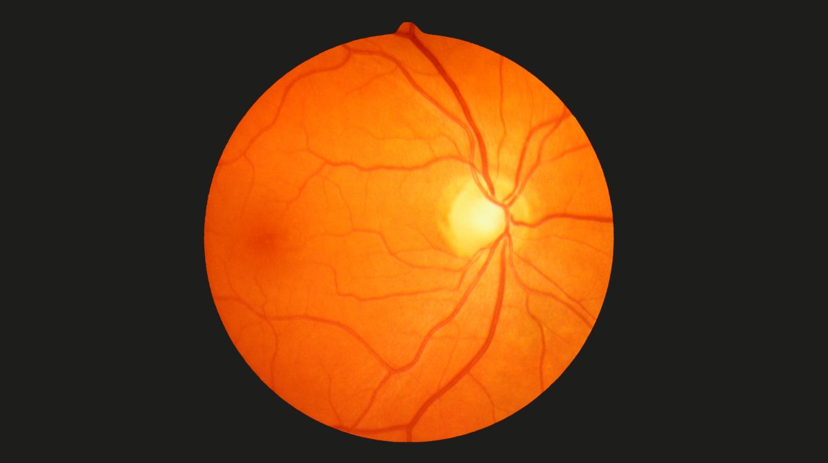
A study conducted on older people shows that changes in retinal vascularization observed during fundus examination are associated with the occurrence of dementia. Therefore, this easily accessible screening could help to identify those affected at an early stage in order to offer them care adapted to this risk.
Our blood vessels, especially the small caliber ones, can deteriorate as we age. At the cerebral level, vascular lesions are therefore associated with most dementia in older people, even if these cognitive disorders are of neurodegenerative origin. However, certain age-related pathologies share common mechanisms and interact with each other. This would be particularly the case with ophthalmological and neurological diseases. Hence the idea evaluated by Catherine Helmer, research director Insertand his Bordeaux team undergo a retinal scan to identify early people at highest risk of dementia.
500 people followed for 10 years
The epidemiological and clinical connections between eye diseases and brain diseases are the focus of the laboratory in which the researcher works. “ The association between retinal changes and small vessel damage in people with dementia is well known, but no long-term study has been conducted to evaluate it Parameters of the retinal vascular network in relation to the occurrence of later dementia have not yet been recorded, she specifies. However, if we confirm that vascular abnormalities at the eye level reflect those at the brain level, we could consider using ophthalmologic examination, which is much faster and less expensive than brain imaging, to detect people most at risk of dementia. » The researcher filled this gap by analyzing data from more than 500 people aged 72 or older who were followed for 10 years as part of the studyStudy of the Three Cities (3C). When these people were recruited, they had no cognitive problems. They took part in cognitive and neurological tests and various examinations, including non-invasive imaging of the retina (fundus). “ Our analysis shows that participants who had increased arterial tortuosity in the retina had a higher risk of developing dementia in the following decade. Similar associations have also been observed between vein diameter and the development of mixed or vascular dementia. »
Optimizing care to reduce cognitive impairment
Observing abnormalities in retinal microvascularization could therefore really help identify people at risk of dementia, although this association remains to be confirmed in other cohorts. The researcher would also like to complement these results with other more advanced retinal imaging techniques, such as: tomography through optical coherence (or OCT angiography), which allows imaging the retina in three dimensions and with much higher resolution on the capillary scale. To find out whether even earlier identification would be possible, she plans to subsequently carry out this work on younger people. Ultimately the idea would be, “ suggest as soon as possible a “Optimized care – taking into account all factors that promote cognitive impairment, such as high blood pressure, diet or lack of exercise, etc. – for people at risk of developing age-related dementia.”
Catherine Helmer is Inserm’s research director on the team Exhibitions on the topic of “whole life, health, aging” (LEHA). under the direction of Cécile Delcourt at the Research Center for Population Health in Bordeaux (BPH, Unit 1219 Inserm/University of Bordeaux).
source : SC Lima Rebouças et al. Retinal microvasculature and incident dementia over 10 years: The Three City Alienor cohort. Alzheimer’s Dementia (Amst), October 22, 2023; doi:10.1002/dad2.12480
Author: CG
also read