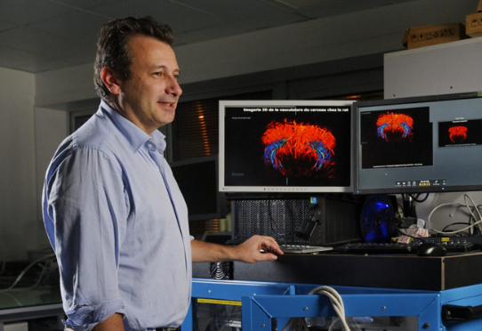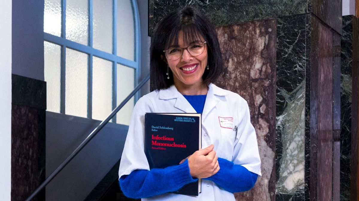
Winner of European funding, Clément Papadacci, physicist and researcher Insert, is developing an ultra-fast ultrasound probe that can be used to observe microvessels of entire organs in three dimensions. Its first applications will be dedicated to the study of neurological and cardiac diseases, which have previously been difficult to study in detail.
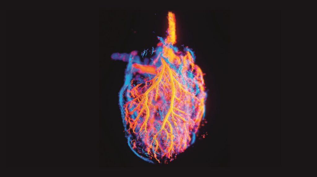
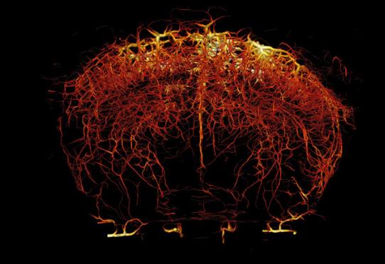
Clément Papadacci has just developed a new type of ultrasound probe: thanks to its high technicality and its dimensions (10 cm x 10 cm), it allows obtaining a large image, a sensitivity and a resolution unlike those of traditional ultrasound or other imaging methods lies . This three-dimensional imaging is carried out after the injection of a contrast agent made of stable and biocompatible microbubbles and is intended to make it possible to visualize the finest vessels (with a diameter of approximately 0.1 mm) on an organ scale. Whole like the heart or the brain. In order to realize his development, the researcher received funding from the European Research Council (ERC Starting Grant) in 2022.
Observe the unexplored
Clément Papadacci is already expecting the first clinical and preclinical applications. There are two diseases in the viewfinder at the moment. “ THE Glioblastoma, a rare brain tumor, must be nourished by microvessels in the blood to grow. We believe that ultrafast ultrasound imaging could help identify these vessels and thus early predict recurrences that occur after surgery. » In the field of cardiology, however, the researcher would like to deal with the microvascular dysfunction of the coronary arteries: This damage to the small blood vessels of the heart, which can lead to angina pectoris-like chest pain, cannot be observed using current imaging methods: “ By giving doctors a first opportunity to examine closely, we could support the development of targeted treatments. »
More broadly, the probe developed by Clément Papadacci and his team could represent a technological turning point in the field of imaging. “ It is expected that this technology will attract widespread interest in the field of ultrasound. European funding enables us to position ourselves as a pioneer in strong international competition. We have to maintain this lead! » Today, the researcher is working on the actual construction of this probe as well as optimizing the algorithms for recording and using the collected data. “ It is a unique opportunity to receive such funding and only in Europe do we dedicate so many resources to a project like MicroflowLifeemerged from concepts in fundamental physics, wave physics and optics. »
At the interface of physics and medicine
A collection of disciplines familiar to this physicist and Inserm research fellow at the Physics for Medicine Institute of the School of Industrial Physics and Chemistry of the City of Paris. It is deeply rooted in the scientific history of this institution “Cradle of ultrafast imaging” that he began his scientific career with his doctorate. “ It’s an environment with Strong dynamism and a real vision at the interface between physics research and its medical applications. The laboratory’s researchers carry out many joint projects: there is a real team culture that is very stimulating. But this is one of the key elements of good research. »
When his path seems to be paved with success, ” nothing is ever acquired,” warns Clément Papadacci. The difficulty lies in the job: “ We are constantly experiencing an emotional rollercoaster, especially when our results do not meet our expectations. But every difficulty is an opportunity to test our theories and progress. ” A job ” sometimes unpleasant ” Yes but “ always creative », which he decided on after acquiring two licenses at the same time, one in physics, the other in cinema! “ From my first internships in the laboratory, I knew that research would give me the opportunity to combine the rigor and complexity of the fundamental discipline with the creativity I was looking for. » And MicroflowLife could well be one of the future milestones in medical imaging.
Clément Papadacci is a researcher at the institute Physics for medicine (Unit 1273 Inserm/CNRS/ESPCI/Paris-PSL), in Paris.
Author: CG
also read